|
|
 |
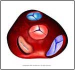
Normal |
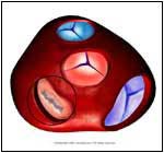
Abnormal |
| |
- The heart has four chambers, two on the left and two on the right. The upper chambers are known as the atria, and the lower chambers are known as the ventricles. The mitral valve, which has two leaflike cusps, acts like a door that regulates blood flow between the left atrium and the left ventricle. The normal direction of blood flow is from the atrium to the ventricle (when the mitral valve is open). When the mitral valve closes, the blood is not allowed to flow back to the atrium (i.e., regurgitate). When the mitral valve is damaged as in Mitral Regurgitation (MR), the normal direction of flow is compromised, and various symptoms develop.
|
 |
- May be symptomatic for years
- As the years go by, the MR causes the ventricle to enlarge, causing Heart Failure. The Symptoms of Heart Failure are:
- Shortness of breath from normal
activities, such as walking or climbing stairs.
- The inability to lie flat without
getting short of breath (may need 2-4 pillows at night).
- Suddenly awaking at night with a
shortness of breath
- Ankle swelling
- Fatigue, weakness, and weight
loss.
- With long term MR, the atrium
enlarges, causing irregularities in the heart rate (such
as Atrial Fibrillation).
- Blood clots in the atrium may be seen
|
 |
- Rheumatic heart disease -- a childhood infection with Streptococcus (strep-throat) can cause Rheumatic Fever,
which damages the heart valves.
- Damage to the mitral valve due to:
- Myxoma -- A form of heart tumor
- Coronary heart disease -- as the
blood vessels of the heart are blocked due to disease, the
oxygen supply to the Mitral valve is compromised.
- Infections of the blood that damage
the valves.
- Calcium crystals can
form on the valves.
- Mitral valve prolapse (MVP) happens
when the chords that hold up the mitral valve are damaged.
- Connective tissue disorders are a group of diseases (such as Marfan's Syndrome) that can damage the Mitral valve.
|
 |
- Medical history (i.e., of Rheumatic Fever etc.)
- Medical exam -- the physician can hear the regurgitation (a murmur). There may also be other signs if the heart is enlarged and failing.
- A chest X-Ray may reveal Fluid in the Lungs and heart, which causes enlargement to occur.
- An Electrocardiogram (EKG), which gives you information of the electrical flow in the heart, may show ventricular enlargement and abnormal rhythms (atrial fibrillation).
- An Echocardiogram uses
sound waves to show the flow of blood across the Mitral valve. One can also see the condition of the valve (i.e., Calcium deposits, bacterial growth, tumors, and ruptured chords), and assess the function and size of the chambers.
- Cardiac catheterization and Ventriculography are tests done in the hospital by a cardiologist. These techniques involve injecting dyes (colored chemicals) into the veins (often the femoral vein in the groin area), and taking X-Rays.
|
 |
- Untreated strep throat
- Coronary Heart Disease
- Intravenous drug abuse (i.e., shooting heroin into the veins with a dirty needle).
- Females have higher rate of MVP
|
 |
- Medications to reduce fluid overload (DIURETICS)
- Digitalis improves the pumping ability of the heart, normalizing an abnormal heart rate.
- Other medications such as nitrates and Capoten help to relieve some of the pressure on the heart.
- Follow up with a cardiologist (a heart specialist) every 3-6 months.
- Surgical repair or replacement of the valve can be done.
- With an enlarged atrium, blood thinners (Warfarin) are given to stop the formation of clots.
- Before any surgical or dental procedures are performed, a patient with MR must be given antibiotics to reduce the risk (Prophylaxis) of Mitral valve infection.
- Have your physician examine you thoroughly. Information from the American Heart Institute is available by calling 1-800-242-8721.
|
| | |
If you want your friend to read or know about this article, Click here
 |
|
|