|
|
 |
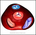
Normal |
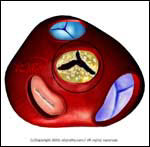
Abnormal |
| |
- The heart pumps blood from its left ventricle (left
lower chamber) to the rest of the body by way of a
large blood vessel known as the aorta. The aortic valve,
located between the left ventricle and the aorta, opens when
the ventricle pumps blood to the aorta, and closes
(passively) when at rest (i.e., between heartbeats).
Normally, the aortic valve has three leaflets.
- If the valve becomes narrowed, it causes Aortic Stenosis,
interfering with the heart's ability to pump blood to the
rest of the body. (Think of a hose blasting water through a
crimped opening).
- Aortic valvular stenosis is due to the progressive buildup of Calcium on the valve leaflets, or when the valve leaflets suffer damage. (Note: severe Aortic Stenosis is
defined as a valve area of 0.7 square centimeters or
less.)
|
 |
- Shortness of breath
- Lightheadedness especially on
exertion
- Fainting on standing or exertion
- Chest pain
- Rarely, sudden
death
|
 |
- Congenital bicuspid valve -- the aortic valve has two leaflets instead of the normal three, causing Calcium buildup and
progressive valve constriction.
- Rheumatic heart disease -- caused by
untreated "strep throat" infections usually from childhood
- Elderly individuals (without specific
cause)
|
 |
- Carotid -- delayed and diminished
carotid upstroke
- Heart in mild to moderate cases
will reveal a systolic eject murmur in the aortic area
that radiates to the neck and apex
- In severe cases -- reversed
splitting of the second heart sound or weak/absent aortic
sound. Signs of left ventricular hypertrophy may be
present, such as left ventricular heave or thrill.
- Lungs -- signs of Heart Failure may occur in severe Aortic Stenosis
(e.g., crackles)
- Electrocardiogram
may show left ventricular hypertrophy, repolarization
changes, or may be normal
- Chest X-Ray may show a
calcified aortic valve and cardiomegaly
- Echocardiogram can
evaluate the valve and the degree of stenosis (when done
with a Doppler)
- Cardiac catheterization gives the
definitive measurements of
stenosis.
|
 |
- Aortic Stenosis is
treated by surgical valve replacement when it causes
symptoms, or when stenosis (narrowing) becomes severe.
- The valve may be replaced with a mechanical (artificial valve) or porcine (pig) valve. Mechanical valves may be more durable, but require anticoagulation with the blood thinner Coumadin. A new procedure involves transplanting the patient's own pulmonary valve to the aortic area, and replacing the pulmonary valve instead. (Since the aortic valve is the one under greater pressure, a transplant as described above will lower the risk of rejection and decrease the need for repeat replacement surgery.) Prior to surgery, the patient is placed on a low Sodium diet, diuretics
("water pills"), and Digoxin.
- Balloon angioplasty (opening a
balloon device in the stenotic valve to open it) is used
primarily in patients for whom surgery is not an option, or
as an alternative to
surgery.
|
 |
- See your physician as soon as
possible. If you have symptoms such as chest pain, shortness
of breath, or fainting seek immediate emergency medical
treatment.
|
 |
- Patients with Aortic Stenosis or
those who have had valve replacement should be placed on antibiotic prophylaxis to prevent infective endocarditis. This includes dental, respiratory, esophageal, gastrointestinal, and genitourinary procedures.
|
| | |
If you want your friend to read or know about this article, Click here
 |
|
|